The Morphology and Function of our Skin as Defence and its Reported Manifestations with COVID-19
- Australian Society Dermal Clinicians

- Mar 5, 2021
- 7 min read
PART 1: OUR SKIN
Lets first introduce our physical barrier between the outside and inside human environment.
The epidermis is known to serve out its primary role of protection through its composition of various layers and constituents. The composition and properties of the most superficial epidermal layer, the stratum corneum, are responsible for its function as a barrier to external influences. The “bricks and mortar” combination of protein-rich layers of terminally differentiated corneocytes within a continuous lipid matrix provides the barrier function of the stratum corneum. An indication of a healthy skin is the optimal efficacy of the epidermal barrier, providing protection against external pathogens, physical and chemical assaults and ultraviolet radiation; while also allowing regulated movement of water and electrolytes to maintain homeostasis. Barrier function is affected by various external and internal influences, such as disease, age, ethnicity, diet, seasonal variations and environment, physiological
stress which leads to an increased level of glucocorticoids, and abnormal enzyme activity. Barrier properties also vary depending on anatomical location, thickness of the stratum corneum, concentration of skin appendages and stratum corneum hydration level.
The morphology at the basement membrane begins as a row of simple cuboidal keratinocytes, and then as differentiation occurs, cells accumulate more keratin and
their morphology alters to its final form of stratified squamous epithelium at the stratum corneum. The morphology of the basement membrane cells gives rise to its function in providing nutrients from its adjacent dermal connective tissues to its neighbouring, avascular epidermal layers above. The simple layer of cells allows this exchange to occur promptly and efficiently. Additionally, their cuboidal shape allows room for a large nucleus and organelles to ensure mitosis can occur continuously. Frequent replication is a unique and protective feature of the epidermis.
A squamous morphology denotes flat, thin cells that can be tightly packed. The cells
are de-nucleated, keratin rich, plasma membrane enclosed bundles functioning
primarily in rapid transmembrane movement. This morphology gives the cells an
ability to regulate various substances passing into deeper strata, serving to protect
the underlying tissue from non-desirable substances. The stratification of these
upper cells means there are multiple layers present to form a dense, compact sheet
filled with tough, fibrous, intracellular protein called Keratin. Keratin alone protects
the underlying tissues from heat, microbes, chemicals and abrasions.
The epidermis is also abundant in covalently-bound lipids such as ceramides, fatty
acids and cholesterol organised into lamellar bilayers, which serve to retain or repel
water, and cellular junctions such as desmosomes to provide cellular strength and
resilience. The extra-long chains of the stratum corneum ceramides contribute to its barrier function. Lipids also function to signal cell proliferation and programmed cell death. Linoleic acid, the primary fatty acid in the stratum corneum, also ensures the barrier quality of the epidermis. Hyaluronic acid present in the epidermis may also contribute to stratum corneum barrier function and hydration. Small amounts of cholesterol esters and cholesterol sulfate within the stratum corneum lipids are also vital to optimal barrier function. Lipids are organised as a lamellar bilayer, providing the essential element as a water barrier. Surrounding the corneocytes, the cornified envelope consists of crosslinked proteins filaggrin, loricrin and involucrin, and provides the physical barrier properties. Additionally, various cells such as the Langerhans cell and Melanocyte provide protection through foreign body removal and DNA damage prevention respectively.
Within the epidermis are three elements of the skin barrier which act together as a
coordinated first line of defence. The stratum corneum serves as the air-liquid interface barrier, tight junctions form the liquid-liquid interface barrier, and Langerhans cell network forms the immunological barrier. The weakly acidic nature (pH 4.0 – 6.0) of the stratum corneum is a self defence mechanism against invading pathogens, as is the presence of commensal bacteria as an element of the innate immune system.
Keratinocytes within each of the four epidermal layers display characteristics of the
sequential differentiation stages. Cells undergo proliferation then progressive differentiation as they upwardly migrate through the epidermis. Cornification describes the conversion of viable epithelial cells to denucleated but still functional corneocytes. Cornification consists of three events; formation of the intracellular keratin network, establishment of the cornified envelope, and formation of the intercellular lipids secreted from lamellar body contents. The corneocytes, encapsulated within the tough cornified envelope and joined by corneodesmosomes, protect against physical and chemical abrasion, maintaining the structural integrity of the stratum corneum. Corneodesmosomes undergo gradual enzymatic deterioration and replacement through the process of desquamation, or shedding of superficial corneocytes.
The stratum corneum is a highly specialised structure, a sophisticated biosensor that
sends signals to the underlying epidermis to respond to external stimuli. Depending
on the desired outcome, the barrier properties of the epidermis are either advantageous or detrimental. For example, substances that are intended to penetrate the skin can do so via intercellular (between stratum corneum layers), transcellular (through keratinocytes), or transappendageal pathways (through sweat glands, sudoriferous glands or hair follicles.)
A damaged barrier ideally results in re-epithelialisation by migration of epidermal
keratinocytes from nearby follicles. The wound healing response of the epidermis
and the pigmentary changes following UV exposure are visible skin manifestations
of the constant endeavor to maintain homeostasis.
Our skin is a dynamic, flexible, selectively permeable barrier with a complex structural organisation, designed as the interface between the internal and external environment. The keratinised stratified squamous epithelium suits tissues with a protective function, such as the epidermis. Cell renewal and replacement is usually a well-organised, effective process which maintains a constant barrier and therefore, homeostasis. Stratum corneum corneocytes embedded in a lipid matrix, likened to a bricks and mortar structure, provide this effective barrier. Defects in the above constituents could lead to an impaired barrier, leaving the skin vulnerable to pathogens. The function or dysfunction of the stratum corneum therefore determines the quality of the barrier.
PART 2: COVID-19 and Documented Skin Manifestations
COVID-19, caused by the novel coronavirus, SARS-CoV-2, was initially identified in
Wuhan, China in 2019. The virus spread rapidly until it was declared a global
pandemic by the World Health Organisation (WHO), with the most commonly
reported symptoms including fever, dry cough, sore throat, headache, asthenia,
myalgia and breathing difficulties. According to The Australian Government
Department of Health it is strongly emphasised that through the practice of good
hygiene and social/physical distancing the spread of COVID-19 can be decelerated.
Yet, as the number of COVID-19 cases develops worldwide, so does our knowledge
and understanding of its presenting manifestations. Currently, there is a mass of
published evidence indicating various dermatological signs of COVID-19. This is
unsurprising considering the skin operates as a protective barrier through its own
unique immune system with many skin disorders and manifestations signifying an
immune response in process.
Presently, the cutaneous manifestations associated with COVID-19 are generally
unknown and are regarded as sporadic presentations of the virus. But case reports
and case series are rapidly accruing and are hence being documented in the
literature. It is understood that the associated cutaneous manifestations are being
unidentified due to the shortage of dermatology consultations in this patient group.
Based on the recorded evidence and data to date, it is speculated the cutaneous
manifestations linked with COVID-19 are parallel to those of other known common
viral infections.
Currently, the published dermatological signs of COVID-19 include:
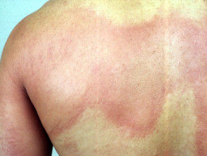
Erythematous exanthems: This is a widespread rash on the trunk and extremities that is usually resultant of infectious anomaly’s, such as a virus. This appears as spots or blotches and may be itchy.

Urticaria: This is characterised by weals, also known as hives and/or angioedema, or swelling.
Weals are superficial skin coloured swellings usually surrounded by redness. They may be accompanied by a burning sensation and may be itchy.
Angioedema is a deeper swelling within the skin or the mucous membranes and can present as skin coloured or red. These may be itchy and painful.
Vesicular or bullous lesions: These are fluid filled lesions, much like a blister.

Chilblains: These are itchy and tender red or purple bumps.

Livedo reticularis: This is a mottled discolouration of the skin.
Vasculitic lesions: These are inflamed blood vessels in the skin and present as purpura, or blood spots.

Petechiae: These are small purpuric lesions
Acrosyndromes (erythromelalgia, raynauds): Erythromelalgia is characterised by an intense burning pain and redness. There is also an increased skin temperature, primarily of the feet and the hands.

Raynauds is an episodic reduction in the blood supply to the fingers and/or toes.

Burning: A generalised burning sensation of the skin.
With respect to the abovementioned, it is essential to note that there is no present
evidence to indicate the extent of cutaneous involvement is linked with the severity of
COVID-19.
Whether dermatological manifestations observed in patients with COVID-19 are
interrelated with the virus itself remains elusive. The skin outbreaks presenting in
COVID-19 patients could also be consequential of other viral infections, systemic
results or a side-effect of prescription medications. Though, clinicians should occupy
a high index of scepticism and ensure they are equipped for patients presenting with
cutaneous eruptions secondary to the COVID-19 virus. Also, clinicians must
understand that early detection of cutaneous signs in combination with swift
management is fundamental in improving patient outcomes.
References
The Australasian College of Dermatologists. (2020). Dermatological manifestations
of COVID-19.
Coronavirus (COVID-19) health alert. (2020, April 27). Australian Government
Department of Health. https://www.health.gov.au/news/health-alerts/novel-
coronavirus-2019-ncov-health-alert
(2017). DermNet NZ – All about the skin | DermNet NZ. https://dermnetnz.org/
Landa, N., Mendieta‐Eckert, M., Fonda‐Pascual, P., & Aguirre, T. (2020).
Chilblain‐like lesions on feet and hands during the COVID‐19 pandemic.
International Journal of Dermatology. https://doi.org/10.1111/ijd.14937
Morey-Olivé, M., Espiau, M., Mercadal-Hally, M., Lera-Carballo, E., & García-
Patos, V. (2020). Cutaneous manifestations in the current pandemic of coronavirus infection disease (COVID 2019). Anales de Pediatría (English Edition). https://doi.org/10.1016/j.anpede.2020.04.002
Recalcati, S. (2020). Cutaneous manifestations in COVID-19: A first perspective. Journal of the European Academy of Dermatology and Venereology. https://doi.org/10.1111/jdv.16387
Rivera-Oyola, R., Koschitzky, M., Printy, R., Liu, S., Stanger, R., Golant, A., &
Lebwohl, M. (2020). Dermatologic findings in two patients with COVID-19. JAAD Case Reports. https://doi.org/10.1016/j.jdcr.2020.04.027
Suchonwanit, P., Leerunyakul, K., & Kositkuljorn, C. (2020). Cutaneous
manifestations in COVID-19: Lessons learned from current evidence. Journal
of the American Academy of Dermatology. https://doi.org/10.1016/j.jaad.2020.04.094
Boer, M., Duchnik, E., Maleszka, R., & Marchlewicz, M. (2016). Structural and
biophysical characteristics of human skin in maintaining proper epidermal barrier
function. Advances in Dermatology and Allergology, 1, 1-
Bäsler, K., Bergmann, S., Heisig, M., Naegel, A., Zorn-Kruppa, M., & Brandner, J. M.
(2016). The role of tight junctions in skin barrier function and dermal
absorption. Journal of Controlled Release, 242, 105-118. https://doi.org/10.1016/j.jconrel.2016.08.007
Dąbrowska, A., Spano, F., Derler, S., Adlhart, C., Spencer, N., & Rossi, R. (2017). The
relationship between skin function, barrier properties, and body-dependent
factors. Skin Research and Technology, 24(2), 165-
Matsui, T., & Amagai, M. (2015). Dissecting the formation, structure and barrier
function of the stratum corneum. International Immunology, 27(6), 269-
Menon, G. K., Cleary, G. W., & Lane, M. E. (2012). The structure and function of the
stratum corneum. International Journal of Pharmaceutics, 435, 3-
Osseiran, S., Cruz, J. D., Jeong, S., Wang, H., Fthenakis, C., & Evans, C. L. (2018).
Characterizing stratum corneum structure, barrier function, and chemical content
of human skin with coherent Raman scattering imaging. Biomedical Optics
Express, 9(12), 6425. https://doi.org/10.1364/boe.9.006425
Pullar, J. M., Carr, A. C., & Vissers, C. M. (2017). The roles of vitamin C in skin
health. Nutrients, 9(8), 866. https://doi.org/10.3390/nu9080866
Ramos-e-Silva, M., & Jacques, C. D. (2012). Epidermal barrier function and systemic
diseases. Clinics in Dermatology, 30(3), 277-
Tortora, G. J. (2018). Principles of anatomy and physiology (2nd ed.).
Van Smeden, J., Janssens, M., Gooris, G., & Bouwstra, J. (2014). The important role of
stratum corneum lipids for the cutaneous barrier function. Biochimica et
Biophysica Acta (BBA) - Molecular and Cell Biology of Lipids, 1841(3), 295-313. https://doi.org/10.1016/j.bbalip.2013.11.006


